版權:新思惟國際
第一篇下載 / 第二篇下載
指定論文說明
《個案報告、技術發表與文獻回顧工作坊》的指定論文比較特別,總共有兩篇。

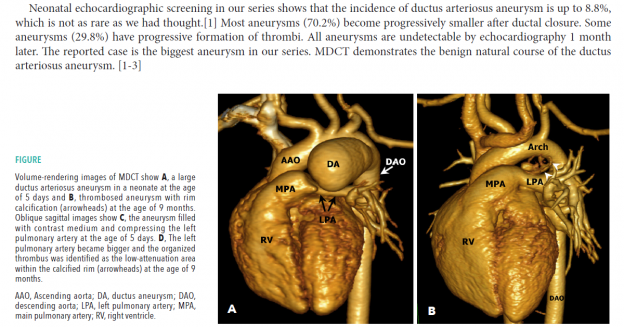
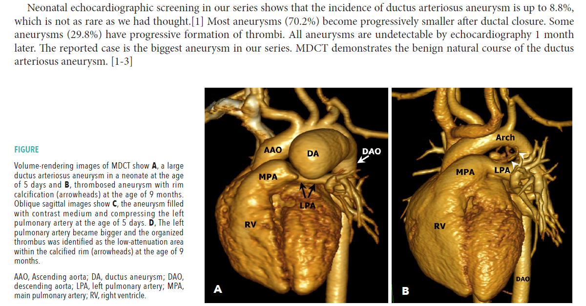
第一篇是刊登在兒科頂尖綜合雜誌 Journal of Pediatrics 的 case report,蔡依橙與傅雲慶醫師團隊,介紹剛出生時發現的巨大 ductus arteriosus aneurysm,在一個月內幾乎完全自行消退的案例,以優質的圖片以及簡要的概念陳述取勝。
- Tsai IC, Fu YC, Jan SL, Lin MC, Ho CL, Hwang B. Spontaneous regression of a large ductus arteriosus aneurysm in a neonate. J Pediatr. 2008 Jul;153(1):143.

第二篇則是刊登在放射科頂尖綜合雜誌 American Journal of Roentgenology 的 review,內容是蔡依橙校長團隊施作 CT-guided biopsy 的完整技術介紹,也是 technical note。目前這篇在 Google Scholar 上的引用次數已經破百。
內容包括操作技術、施作現場、關鍵步驟說明,並附上許多動畫,值得參考。請搭配本頁下方的 GIF 動畫與說明閱讀。
- Tsai IC, Tsai WL, Chen MC, Chang GC, Tzeng WS, Chan SW, Chen CC. CT-guided core biopsy of lung lesions: a primer. AJR Am J Roentgenol. 2009 Nov;193(5):1228-35.

新思惟國際,尊重各種形式的智慧財。所以,我們取得蔡依橙與傅雲慶醫師同意後,拿到論文的 MS-Word 版本原稿重新編輯,也委請設計師,用不同的風格,重新排版過。版權由「新思惟國際」回贈給蔡依橙醫師與傅雲慶醫師,並均能獨立使用。
推薦的閱讀方法,是一邊閱讀時,一邊思考,我手上是否有保留這樣的 case 可以投稿?我的技術是否有足夠的案例與廣度,能寫成這樣的技術回顧,並讓期刊社認為有教育意義值得刊登?我的臨床案例收集流程,是否能讓我把握住這樣的機會?我的圖片處理能力,是否能到達這樣的程度?
一有問題,就在旁邊寫下來,歡迎您在課堂上,隨時與原作者詢問討論。讓自己成為有能力產出這兩篇文章的高手。
Animations for AJR CT-guided lung biopsy article
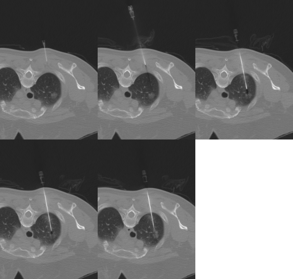
Animation 1 – The dynamic process of CT-guided lung biopsy for a 57-year-old female’s 1.5-cm right apical lung nodule, later proven to be an adenocarcinoma.
After local anesthesia, the hypodermic needle was left in the chest wall with syringe removed to see the direction of planning. Then the coaxial needle was inserted into the chest wall, through the lung parenchyma and to the periphery of the tumor with several attempts at manipulation. Because the needle inserted is restricted by the skin, muscle and even lung parenchyma, the deeper the needle, the larger the manipulating angle needed for any adjustment. While adjusting the needle, avoid pulling back and puncturing the pleura multiple times. The core biopsy was performed, causing small parenchyma hemorrhages in the regions surrounding the coaxial needle. In the animation, all images are in the same position. The patient was also cooperating well, but still obviously moving, especially when the procedure involved some pain. Thus, the dynamic process means we must adjust the needle according to the latest scan and the relationship between the inserted needle and the tumor, rather than strictly following the initial plan. The needle insertion process is not a one-step procedure, since most cases need several manipulation attempts to reach the tumor periphery.
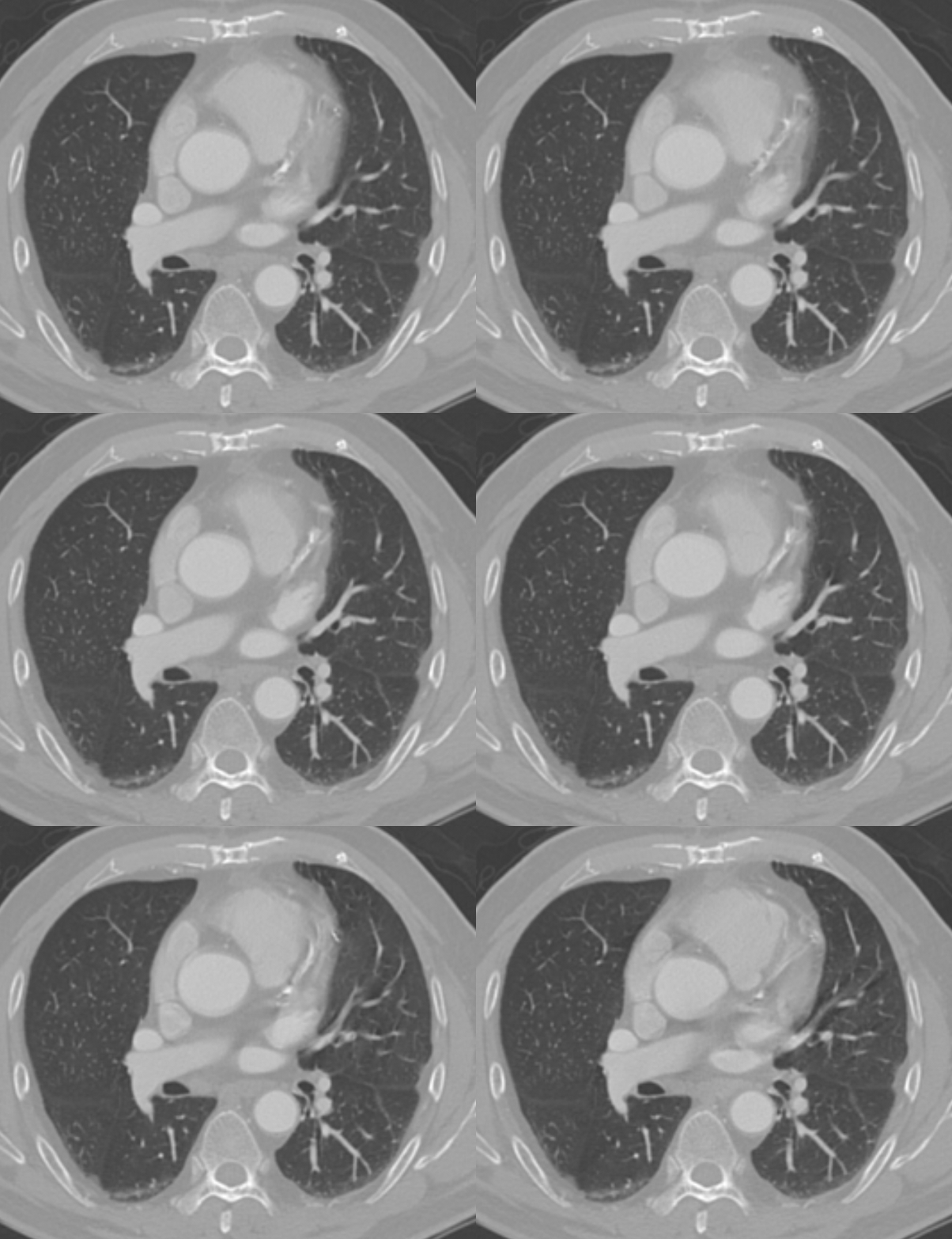
Animation 4a – Lung motion during heart cycle at the level of aortic root.
The motion is obvious near the right ventricular outflow tract, the right atrium and the superior aspect of the left ventricle. Please notice the vigorous movement of the lung parenchymal vessels in the left lingula. The right middle lobe is also affected by the cardiac motion caused by the right ventricular outflow tract, right atrium and right ventricle, but to a lesser extent.
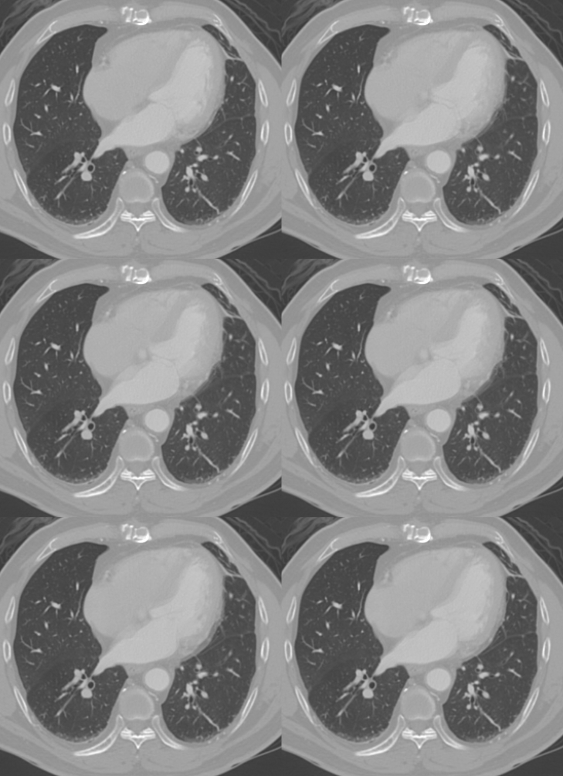
Animation 4b – Lung motion during heart cycle at the level of left ventricular outflow tract.
The left lingula is affected by left ventricular motion. The motion over the left ventricular lateral wall is about 1 cm. Avoid biopsy in regions showing motion artifact. At this level, the right middle lobe is not much affected by right ventricular motion.
最新活動
